Introduction
The skin has been described as the largest organ in
the body. It defends the body it covers and is involved
in the maintenance of homeostasis including
water conservation. The skin is involved in body
temperature conservation through insulation and in
heat loss through perspiration. The sensory nerves of
the skin recognise pain and temperature extremes.
The skin provides protection against minor physical
injuries, supports hair growth and offers some
defence against microbial invasion.
The condition of the skin is a reflection of the
general health of the animal, deteriorating in cases
of ill health, ill thrift and debility. In some conditions,
such as jaundice, the skin may provide through
discolouration direct diagnostic evidence of a specific
disease process. In other conditions, such as
parasitism or severe mineral deficiency, a nonspecific
general deterioration of skin health may
occur causing a greater number of hairs than normal
to enter the telogen or resting phase and a delay in
their replacement, leaving the coat in poor condition
with little hair. Sebaceous secretions may be reduced,
allowing the skin to become abnormally dry and inflexible
and less able to perform its normal defence
role in an already debilitated animal. In other cases
sebaceous secretion increases causing the skin to
have either a greasy or a dry seborrhoeic, flaky
appearance.
The mutual dependency of the skin and the body it
covers must be borne in mind during every clinical
examination. Abnormalities of the skin may be
caused by specific skin disease or by the poor general
health status of the patient. Adetailed clinical examination
of the patient and of its skin are essential parts
of the process of diagnosis and should enable the
health status of the patient’s body and its skin to be
determined.
Applied anatomy
The skin has three main layers: the epidermis,
dermis and subcutis. The epidermis consists largely
of epithelial cells and pigment. The epithelial cells
of this layer are produced by the stratum germinativum
and as further cells are produced reach the
outer surface of the skin in about 3 weeks. Here they
become keratinised, die and are lost from the skin as
a result of contact with the animal’s environment.
The dermis is a connective tissue layer containing
blood vessels, nerves, hair follicles, sebaceous and
sweat glands. The subcutis contains fibrous and
fatty tissues which provide insulation for the body
and support for the outer skin layers. The skin has
considerable elasticity in the normal animal, allowing
body movements to occur. This elasticity may
be reduced by ill health, especially in dehydrated
animals, and also as a result of inflammation and
injury to the skin.
Hair follicles cover much of the bovine body but
are not present at the mucocutaneous junctions or the
surfaces of the muzzle and teats. Most cattle shed
part of their coats in the spring. Considerable hair
growth occurs as cold weather approaches in the
autumn.
History of the case
The general history of the case will have been considered
at an earlier stage in the process of diagnosis.
There are specific points of history, however, that
may have direct bearing on the consideration of skin
disease. The history of the herd and a knowledge of the
geographical area may provide useful information
for the clinician. In areas where copper deficiency occurs,
changes in coat colour may be seen. Previous
skin disease problems on the farm with details of
their diagnosis and treatment may provide a useful
background of information which will assist in the
evaluation of the present case.
The history of the patient, including recent contacts
with other cattle at shows or markets, may also be
important. Recent changes in diet and management
should be noted. Poor nutrition can give rise to a dull,
dry, thin and brittle coat. Loss of condition may have
contributed to poor skin health which can itself then
lead to further deterioration in the animal’s general
health. Specific points in the history of the patient
may be useful. The stockperson may report frequent
rubbing by the animal, suggesting pruritus. Failure
to ensure an adequate supply of minerals and vitamins
can contribute to poor skin health. Details of
previous treatment given and the response to such
treatment may also provide useful information.
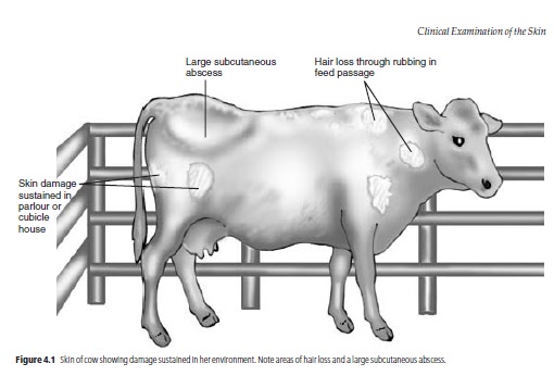
The environment of modern cattle, especially the
dairy cow, contains many features that may damage
the skin. The cubicles, the parlour and the floor may
have abrasive surfaces or sharp corners that can
cause injury to the skin, often repeatedly. Such problems
in the environment are especially likely to be
important if a number of cattle in the herd are seen
with identical superficial injuries. Overcrowding
and insufficient feeding facilities may also contribute
to poor coat condition including superficial skin
damage (Fig. 4.1).
Abnormalities such as a very poor coat, evidence
of excessive self-grooming or large areas of alopecia
may be seen from a distance, but the areas of the
skin must be closely examined too. Opportunities
to examine the skin arise as each part of the body is
examined, but in order to get a general impression
of the skin it can be assessed separately before the
more detailed examination of each area begins.
Visual appraisal of the skin
The whole body surface is methodically inspected
initially from a distance and then more closely, looking
for areas of abnormal skin or hair which will later
be subjected to closer scrutiny. Healthy animals have
lick marks on their skin, especially over the flank
and shoulders. Pruritus, for example that caused by
background of information which will assist in the
evaluation of the present case.
The history of the patient, including recent contacts
with other cattle at shows or markets, may also be
important. Recent changes in diet and management
should be noted. Poor nutrition can give rise to a dull,
dry, thin and brittle coat. Loss of condition may have
contributed to poor skin health which can itself then
lead to further deterioration in the animal’s general
health. Specific points in the history of the patient
may be useful. The stockperson may report frequent
rubbing by the animal, suggesting pruritus. Failure
to ensure an adequate supply of minerals and vitamins
can contribute to poor skin health. Details of
previous treatment given and the response to such
treatment may also provide useful information.
The environment of modern cattle, especially the
dairy cow, contains many features that may damage
the skin. The cubicles, the parlour and the floor may
have abrasive surfaces or sharp corners that can
cause injury to the skin, often repeatedly. Such problems
in the environment are especially likely to be
important if a number of cattle in the herd are seen
with identical superficial injuries. Overcrowding
and insufficient feeding facilities may also contribute
to poor coat condition including superficial skin
damage (Fig. 4.1).
Abnormalities such as a very poor coat, evidence
of excessive self-grooming or large areas of alopecia
may be seen from a distance, but the areas of the
skin must be closely examined too. Opportunities
to examine the skin arise as each part of the body is
examined, but in order to get a general impression
of the skin it can be assessed separately before the
more detailed examination of each area begins.
Visual appraisal of the skin
The whole body surface is methodically inspected
initially from a distance and then more closely, looking
for areas of abnormal skin or hair which will later
be subjected to closer scrutiny. Healthy animals have
lick marks on their skin, especially over the flank
and shoulders. Pruritus, for example that caused by
heavy louse infestation, may cause excessive grooming
and the presence of more lick marks than normal.
Repeated rubbing can lead to hair loss and thickening
of the skin. The presence of any obvious abnormalities,
including swellings or discharging abscesses,
should be noted for further investigation
later. Damp areas caused by sweating may be seen in
pyrexic animals and in warm weather. Skin loss
through injury may be seen. Gangrenous changes in
the skin and deeper tissue may have arisen through
loss of circulation and may be seen or noted during
manual appraisal of the skin.
Manual appraisal of the skin
This should involve as much of the body surface
as possible, using caution when touching sensitive
areas which might cause the animal to kick. Manual
appraisal will enable the clinician to detect lesions
which are not immediately visible, for example beneath
matted hair. Any abnormalities detected are
subjected to further scrutiny which may necessitate
removal of hair and examination of the skin in good
light with the aid of a hand lens. Enlargement of
lymph nodes may be detected at this stage (see
below). The thickness of the skin and the presence of
any subcutaneous oedema or infection should also
be noted. The average skin thickness in adult cattle is
6 mm, with decreasing thickness being evident from
the dorsal to the ventral body surfaces. The skin over
the brisket is quite thick and mobile. This area of skin
may have a spongy texture when compressed and
may give an impression of subcutaneous oedema although
it does not pit on pressure. Genuine oedema
which does pit on pressure may be seen in this area
and between the mandibles in cases of right sided
cardiac failure. The skin covering the lower limbs is
relatively immobile.
Manual examination of the skin will also allow assessment
of skin turgor – its resilience and flexibility.
Picking up a skin fold between finger and thumb and
releasing it provides a general assessment of the animal’s
state of hydration. In a well hydrated animal
the pinched skin falls immediately back into place; in
a dehydrated animal the return to normal is delayed.
The best site for this test is the skin of the upper
eyelid.
Pathological thickening of the skin occurs in a number
of skin conditions, including sarcoptic mange. Thickening
in the form of callus formation can occur in areas
of skin, including those covering joints, which are
repeatedly subjected to trauma. Examples include
the elbows and hocks in animals with poor bedding.
Distribution of skin lesions
This is of diagnostic importance. Lesions caused by
photosensitisation are commonly seen in lightly pigmented
areas on the dorsal parts of the body which
are exposed to sunlight. Such lesions are not normally
seen in pigmented areas. Ringworm lesions
in calves are particularly common on the head and
neck, but also occur elsewhere.
Description of the skin lesions
The clinician should try to determine exactly what
abnormalities are present in the skin, which tissues
are involved and how deeply the disease process
extends into and over the skin. The larger external
parasites such as lice may be seen at this stage. Skin
temperature, thickness, consistency and colour are
observed and compared with adjacent areas. The
presence of subcutaneous oedema or increased skin
turgor is noted: these abnormalities may be caused
by hypoproteinaemia or heart failure and dehydration,
respectively, but they can also be the result of
local pathology. When numbers of skin lesions are
found it is important to determine if they share the
same aetiology. They may represent different stages
of one disease process. More than one condition can
be present at the same time.
There may be abnormalities in the sebaceous and
sweat glands or gross proliferation of the superficial
layers. Self-inflicted trauma can greatly modify and
mask the clinical picture. Skin abnormalities may
involve some or all of the component structures of
the skin: the hair, follicles, epidermal, dermal and
subcutaneous tissues.
Aucun commentaire:
Enregistrer un commentaire