samedi 9 juillet 2011

AUTRES INDICATIONS DERMATOLOGIQUES

Les cortico'ides sont aussi indiqu é s dans de nombreuses dermatoses à médiation
immunologique et aussi néoplasiques. Très souvent, leur prescription souvent longue


nécessite une surveillance thérapeutique étroite. Parmi les nombreuses indications,il convient de citer :
  •la panniculite nodulaire stérile
•la cellulite juvénile pyogranulomateuse,en association avec une antibiothérapie systémique
•la pyodermite pyotraumatique,en association avec une antibiothérapie systémique •les vascularites
•la dermatomyosite,en association avec la pentoxifylline
•le mastocytome multicentrique,le plus souvent associé à d’autres molécules anti-cancéreuses comme la lomustine
•le lymphome cutané T épithéliotrope,aussi associé à d’autres molécules anti-cancéreuses comme l’acitrétine ou la lomustine


Généralement,la posologie d’induction est en moyenne de 1 à 2 mg/kg/jour en une prise pour la prednisone et la prednisolone, et de 0,8 à 1,6 mg/kg/jour en une prise pour la méthylprednisolone. La durée de cette phase d’induction est variable selon les diverses entités (en moyenne plusieurs semaines). Après une phase aboutissant à une phase de stabilisation ou de rémission, le thérapeute adaptera cette posologie en la diminuant et en la prescrivant tous les 2 à 3 jours en fonction de


vendredi 8 juillet 2011

CORTICOTHERAPIE ETDERMATITES AUTO-IMMUNES

Les dermatites auto-immunes (DAI) repr ésentent un ensemble complexe de dermatoses,ayant en commun l’existence de mécanismes d’auto-immunisation vis à vis des composants peau. Il s’agit d’entités rares, causes peu fréquentes de consultation en dermatologie des carnivores domestiques. Leur traitement fait appel à une immunomodulation, potentiellement dangereuse pour l’animal,qui doit toujours être mise en place de façon mesurée et raisonnée.

PRÉALABLES AVANT LA MISE EN PLACE D’UNE
CORTICOTHÉRAPIEPOURLETRAITEMENTD’UNEDAICHEZ LECHIEN

CERTAINES REGLES GÉNÉRALES DOIVENT ETRE SUIVIES :
En premier lieu,il faudra être sûr du diagnostic,ce qui implique une anamnèse et une clinique compatibles, l’élimination des autres hypothèses diagnostiques, et un diagnostic (histopathologique,voire immunologique) de certitude ou fortement évocateur.

En second lieu,le traitement doit être raisonné :il faudra recourir aux drogues les moins toxiques, préférer l’utilisation d’associations médicamenteuses,qui permettent parfois de réduire les effets secondaires à long terme,adapter le traitement en fonction des propriétaires (administration facile, coût...).

En plus du traitement médical,il sera toujours bon d’identifier et d’éviter si possible les causes aggravantes et/ou déclenchantes (DAI photosensibles ou d’origine médicamenteuse).

L’utilisation concomitante de shampooings est toujours intéressante pour réhydrater la peau et éliminer squames et croûtes.
Une récente étude nord-américaine suggère d’utiliser systématiquement une antibiothérapie au début du traitement immunosuppresseur. Cette association permettrait d’augmenter la survie des chiens atteints de pemphigus foliacé.

Il faut également s’assurer de la coopération du propriétaire,et l’impliquer dans la dermatose de son animal :des visites de suivi régulières sont indispensables.

UTILISATION DES CORTICO'iDES EN PRÉSENCE D’UNE DAI
Les glucocorticoïdes sont indiscutablement l’arme de choix en cas de DAI. En effet,ils présentent une action rapide,polyvalente et un coût relativement faible. Leur utilisation doit cependant être raisonnée,car ils peuvent provoquer des effets secondaires,d’autant plus que les doses nécessaires sont souvent importantes et administrées au long cours.
Leur mode d’action dans les DAI est complexe. Ils diminuent la synthèse d’auto-anticorps anormaux,la lymphoblastogenèse,la fonction des neutrophiles,la fabrication du complément et le dépôt des immun-complexes,par baisse de leur passage à travers l’endothélium vasculaire et la membrane basale. Aux posologie utilisées en pratique,leurs effets bénéfiques seraient plus liés à leur action anti-inflammatoire qu’à une réelle immunosuppression.

Les règles générales d’utilisation des glucocorticoïdes sont résumées dans le tableau I. 


On utilisera des substances administrables per os,et qui peuvent être prescrites à jours alternés. La prednisolone et la méthylprednisolone sont les molécules de choix. Les doses d’induction varient : prednisolone (2 à 4 mg/kg/j), méthylprednisolone (1,6 à 3,2 mg/kg/j), dexaméthasone (0,25 à 0,75 mg/kg/j) ou triamcinolone (0,2 à 0,7 mg/kg/j). Les trois premières sont utilisables en jours alternés,les deux dernières ne le sont pas. Ces doses doivent être progressivement réduites dès le contrôle des lésions:on administrera le médicament un jour sur deux pendant une à deux semaines, puis on diminuera la dose de 30 à 50 pour cent tous les quinze jours. Le but est d’atteindre la dose la plus faible possible,administrée le moins souvent possible pour contrôler les signes dermatologiques.

Il faut également envisager le recours en association à d’autres immunomodulateurs, qui permettent au long cours une épargne corticoïde.

EXEMPLE D’UTILISATION : PROTOCOLE DE  TRAITEMENT
DU PEMPHIGUS FOLIACÉ

Le pemphigus foliacé est,après
le lupus érythémateux, la DAI la plus fréquente chez le chien. Nous proposons dans la figure suivante une approche thérapeutique pratique de cette
dermatose.


REMARQUES

La voie parentérale est de peu d’utilité car peu précise,avec des effets variables dans le temps. En outre, les effets secondaires apparaissent
rapidement dans ces maladies chroniques nécessitant
un traitement prolongé.

La corticothérapie pulsée,qui consiste à administrer de fortes doses de corticoïdes par voie intraveineuse pendant de courtes périodes, a été peu étudiée en médecine vétérinaire. Chez l’homme,il ne semble pas qu’elle permette des résultats statistiquement plus
intéressants que les protocoles classiques.
L’utilisation des dermocorticoïdes doit toujours
être envisagée, surtout pour les formes localisées
(exemple : lupus cutané, pemphigus érythémateux),
mais également pour les formes généralisées,
si l’animal est docile. Une récente étude chez l’homme
a en effet montré que l’application de dermocorticoïdes
de classe I sur les lésions bulleuses de pemphigoïde
bulleuse était aussi efficace, aussi rapidement, mais
présentait beaucoup moins d’effets secondaires au long
cours que la corticothérapie par voie générale.

CONCLUSION : SUIVI DU PATIENT

Pour ces dermatites nécessitant une immunomodulation au long cours, des visites de réévaluation fréquentes sont indispensables. Lors de ces visites,le clinicien devra vérifier l’état cutané, et également s’attacher à dépister un début d’intolérance au traitement (bactériurie, polyuropolydipsie,polyphagie,glycémie,…).


jeudi 7 juillet 2011

UTILISATION DES CORTICOIDES DANS LE TRAITEMENT DES OTITES DU CHIEN ET DU CHAT

Les cortico'ides sont le plus souvent utilisés dans le traitement des otites sous la forme de
choisir une stratégie complète de thérapeutique anti-inflammatoire lors d’otite,afin de trouver le juste milieu entre soulagement et facilitation de l’infection.

INTÉRETS ET LIMITES DE LA CORTICOTHÉRAPIE DANS LE
TRAITEMENTDESOTITES


Les corticoïdes agissant à tous les stades de l’inflammation (phases vasculaire,cellulaire et fibrose), ils trouvent leur place dans le traitement de toutes les formes d’otites. L’action sur l’œdème est intéressante parce qu’elle permet de lever l’obstruction canalaire et offre la possibilité de visualiser les conduits dans leur intégralité (et de les nettoyer). L’action antalgique permet de faire plus rapidement des soins locaux et évite les conduites agressives de l’animal. L’action
d’inhibition sur toutes les sécrétions auriculaires est aussi très intéressante parce qu’elle limite les phases de nettoyage et les proliférations infectieuses. Les corticoïdes


UTILISATION DES CORTICOIDES DANS LE TRAITEMENT DES DERMATITES ALLERGIQUES DU CHIEN ET DU CHAT

La corticothérapie a longtemps été le seul outil thérapeutique de la gestion des deratites
 
charge des dermatites prurigineuses du chien et du chat. Afin de limiter ces abus le discours sur le traitement des dermatites allergiques est souvent perçu comme la présentation des moyens pour ne plus faire appel aux corticoïdes. Le traitement des dermatites allergiques ne se limite pas à des attitudes aussi radicales. Il doit être adapté à chaque cas (chaque chien et chaque propriétaire) et les corticoïdes ont une place très importante qu’il faut maîtriser et expliquer. Trop souvent,la prescription des corticoïdes dans le cadre des dermatites allergiques se limite à une approche mixte qui consiste à traiter la poussée puis administrer une corticothérapie à jours alternés, qui ressemble plus à un sevrage qu’à un traitement au long cours. Il est en fait nécessaire de distinguer le traitement de la crise et le traitement au long cours d’une dermatite allergique.

INDICATIONS
La corticothérapie est indiquée dans le traitement de la plupart des dermatites allergiques, à l’exception de certains accidents médicamenteux comme l’érythème polymorphe ou le syndrome de Lyell. Trop souvent,l’abord de la corticothérapie est simplifié et réduit à une posologie de prednisone ou de prednisolone. Or il existe de nombreuses possibilités différentes en termes de posologie,de protocoles thérapeutiques,de molécules ou de galénique.
CHEZ LE CHIEN
 Urticaire et angiœdème
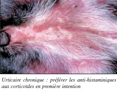
Même s’ils sont souvent prescrits,les corticoïdes ne sont pas indispensables au traitement en première intention des urticaires aiguës. En effet,
ces lésions sont souvent fugaces ou de contrôle aisé avec des anti-histaminiques (anti-H1). Par contre le recours à des corticoïdes d’action rapide est indispensable lors d’angiœdème facial, le risque d’asphyxie par œdème laryngé ou de choc anaphylactique étant toujours possible. Lors d’urticaire chronique,la priorité est donnée à la recherche étiologique, même si elle s’avère décevante dans la majorité des cas. Le traitement


symptomatique repose surtout sur la recherche d’un anti-histaminique efficace.
 DAPP

mardi 5 juillet 2011

Starting Cattle on Feed


I. THE PROGRAM
A recommended program for handling newly purchased feeder cattle upon
their arrival at their new feedlot home is presented. Naturally, it would be
expected that most feedlot operators would plan to impose various modifications
in this program. However, it is quite a workable program when followed in
principle.


samedi 2 juillet 2011

Clinical Examination of the Male Genital System

Introduction
The majority of male cattle are castrated, and in these animals the genital system acts chiefly as a conduit for the passage of urine.
Breeding bulls are an important part of the herd. 
To be effective sires, bulls must be in good health, have a satisfactory conformation and be free, as far as known, from genetic defects.
They should also have a good libido, be able to mount and achieve intromission of the erect penis into the cow’s vagina.
They must be able to ejaculate semen of sufficient quality to fertilise the ova of healthy cows.
In order to establish that a bull has these qualities a comprehensive and methodical examination is necessary.
Applied anatomy


The anatomy of the male genital system is illustrated in (Fig. 11.1). Details of the anatomy of individual genital organs are described below under ‘Clinical examination’. 
Signalment of the case
Details of this can be useful in the diagnosis of male breeding problems.
Although puberty may be reached at 9 months, bulls are generally not used as sires until they are 18 months of age or over.
Younger animals may have a poorly developed libido and poor semen quality.
Excessive use of very young animals may also compromise sperm production.
Libido may fall off in older bulls, and heavy bulls may have physical difficulty in mounting to
serve.
Some strains of Hereford bulls have a poorly developed libido.
History of the case
The general history of the case will have been discussed earlier in the diagnostic process.
The owner should be questioned to determine the nature of any current fertility problem, the origin of the bull and his past performance.
The following questions should be asked:
(1) Has the bull been recently purchased?
If so are details available of his past performance?
Has he ever sired a calf?
When was he last known to have sired a calf?
(2) Has the bull been in good general health? 
Has he lost condition recently?
Have there been any signs of lameness or other orthopaedic abnormality?
(3) Did his herd of origin have any past fertility problems?
What was its health status?
Was the herd known to be free from bovine virus diarrhoea infection and other diseases which may affect fertility?
What is the disease status of his present herd?
(4) Has the bull served cows presented to him individually (in hand) or has he been running with
the herd?
(5) If serving cows individually, has he been frightened in any way?
Are the staff handling him experienced in their work?
(6) How many female animals was he running with?
Is he still with them and if not when was he removed from their group?
Has normal oestrus behaviour been observed in the group? 
Have they been bred from successfully before?
(7) What is the condition of the female animals?
If in poor condition for how long has this been a problem?
Has the cause of their poor condition been investigated?
(8) Has the bull been seen to serve? Was his service behaviour normal?
Were any specific abnormalities during service noticed?
In many cases close observation of service may not have been undertaken and the owner may be unaware of important details of service behaviour.
(9) Has pregnancy diagnosis been undertaken on the animals with which the bull has been running?
What method of pregnancy diagnosis was used and was it capable of diagnosing early pregnancy (at 30 days)?
(10) Have the female animals been exposed subsequently to another bull? Has the other bull bred with them successfully?
Observation of the patient
The bull should be quietly observed to see if any obvious abnormalities which might affect his breeding ability are visible.
Few reproductive problems in the male are visible externally. 
Obvious lameness could affect his ability to serve.
Gross abnormalities in scrotal size or a disparity in testicular size may be visible, but must be confirmed and investigated during the full clinical examination.

dimanche 19 juin 2011

Clinical Examination of the Female Genital System


Introduction
Examination and assessment of the genital system is an important part of the veterinary management of dairy cows. 
The target on many dairy farms is for cows to achieve a calving to calving interval of 365
days. 
To achieve this target the reproductive performance of the cow has to be closely managed. Some consider this calving interval to be an unattainable and possibly undesirable goal in high yielding cows.
On many farms it is only achieved by close monitoring of the cows’ reproductive performance and intervention with strategic hormone therapy.
An assessment of herd fertility should involve examination of animals, including any problem animals, as they are presented for routine fertility checks.
It should also involve consideration of the farm husbandry and management. 
Information required should include the overall disease profile of the farm, milk yields and both past and present fertility records.
The cow has an average gestation length of 283 days.
To achieve a calving interval of 365 days she must conceive again within 82 days (365 - 283 = 82) of her previous calving.
Uterine involution is normally complete and resumption of overt ovarian activity has normally commenced by 40 days after calving.
Conception should ideally occur in the period of 40 to 82 days after calving.
Beef cows are subject to less intense pressures because their milk production has to be sufficient only for their own calf. 
None the less, a calving interval of 365 days is very important to enable the herd to calve at approximately the same time and over a short period each year.
Ashort calving period enables the herd and their calves to be fed and managed as a
group.
Monitoring of the reproductive performance is also important in beef cattle.
Much routine fertility work consists of specific and often limited examinations of the cow’s genital system.
A more detailed and comprehensive examination may be requested and is necessary when a
particularly valuable cow fails to conceive.
In every case it must be remembered that the genital system is just one part of the patient. Unless the patient is in good health and her genital system is functioning normally, conception may not occur.
Whenever the genital system is examined the veterinarian should also assess the general health of the patient and be alert to the possibility that disease involving other body systems may also be present.
Applied anatomy The anatomy of the female genital system is illustrated in Fig. 10.1. Details of the anatomy of the individual genital organs are given under clinical examination below.
Signalment of the caseThe age of the patient is very important.
Maiden heifers have never bred and a small proportion may prove unable to do so.
Some may be freemartins being the twin to a male calf and having the genital tract of an intersex.
Other congenital defects resulting in infertility are rare but none the less must be considered in such a group of animals.
Fertility problems tend to increase with the cow’s parity because the risk of acquired abnormalities increases with the birth of each calf.
Maiden heifers have not yet sustained injuries at calving or experienced problems associated with a retained placenta.
These problems are more frequent in older cows. Such animals are more likely to be exposed to dietary deficiencies which can have an adverse effect on fertility.
History of the case 
Details of both herd and individual patient history
are of great importance in the recognition and diagnosis
of problems in the female genital system.
History of the herd
The clinician should seek answers to the following questions:
(1) What is the herd size? Has it increased recently?
(2) Are fertility records for the herd available?
On many dairy farms retrospective computerised records are kept. 
A number of indices of fertility may be available and consideration of them all is beyond the scope of this book.
Among important indices is the herd submission rate which assesses the vital oestrus detection rate of the herd. 
The conception rate and its seasonal change monitored by cumulative sum (Q sum) are also very important. 
Poor fertility indices may indicate overall poor performance. 
They may also indicate that one section of the herd, for example first calf heifers, is performing badly. 
It should also be possible to identify how the herd is performing in the current year compared with previous years.
(3) Herd management – what staff are employed?
Have there been recent changes of staff?
What method(s) of oestrus detection are used?
Is a fertility control scheme in place?
Does it include a postnatal check, prebreeding examination and pregnancy diagnosis? 
What is the herd policy on cows not achieving herd targets such as being in calf by 82 days?
(4) Feeding and production – what feeding regime is used?
Has the diet changed recently?
Are trace element deficiencies known in the area? 
Are metabolic profiles taken from cows to assess feeding and identify deficiencies?
Does the farm have a seasonal policy for milk production?
(5) What is the incidence of herd lameness, metabolic disease and mastitis? 
Have these problems increased recently?
(6) How many cases of abortion occurred during the last year?
Was the cause of abortion diagnosed?
Is a vaccination policy in place?
(7) Is the herd self contained? What was the health
profile of any recently purchased animals?
(8) Is artificial insemination (AI) used? Is DIY AI
used? Are the staff skilled in using AI? Do any
staff members have poor cow AI conception
rates? Is on-farm semen storage satisfactory?
If natural service is used, is the bull known to be
fertile?
History of the cow or heifer
Further questions should be asked or records inspected
to ascertain the following details of the
patient’s history:
(1) The age and parity of the cow.
(2) Has the cow had any previous breeding
problems? What were these? Was treatment
successful?
(3) Details of last calving – date, parturient problems
including dystocia, retention of fetal
membranes.
(4) Dates of observed oestrus since calving – has the
cow cycled regularly? Is she cycling now? Have
her cycles been excessively short or prolonged?
(5) Service details – dates and method of service,
operator, bull or semen used.
(6) Production records of this cow.
(7) Health record of this cow – details of lameness, metabolic disease, mastitis, abortion. Observation of the patient Cows presented for fertility investigation may be confined to a stall or in the parlour.
Wherever possible the cow should be viewed from all sides without restriction, so that her general health and condition can be assessed.
Certain specific changes may be seen which relate to the patient’s reproductive state.
Many of these are normal physiological changes, but the clinician should look carefully for obvious signs of abnormality which can be investigated further at a later stage.
The condition score of the cow should be estimated and confirmed by palpation of the lumbar and sacral regions when the cow is handled.
The score (range 1 = very thin to 5 = obese) has an important influence on fertility.
Cows should have a condition score of 3 at calving, 2.5 when served and 2.5 to 3 when dried off.
The cow in oestrus may appear slightly excitable.
A vaginal discharge of clear tacky mucus (the ‘bulling string’) may be present.
Scuff marks may be seen on her hindquarters and in front of her tuber coxae caused by the feet of other animals mounting her.
Dried saliva from other cows may be seen in similar places. Approximately 48 hours after oestrus the cow may pass a dark red watery vaginal discharge.
Animals suffering from long term cystic ovarian disease may show abnormalities of body shape. Virilism, in which bull-like changes are seen, may occur in animals chronically affected by luteal cysts secreting  progesterone.
Increased development of the neck muscles may occur and the animal may become aggressive.
Chronic exposure to oestrogens produced by follicular cysts may produce signs of nymphomania.
In addition to displaying frequent signs of oestrus, affected animals may show slackening of the
pelvic ligaments with apparent prominence of the tail head.
Adegree of abdominal distension is anticipated during pregnancy, especially in the last trimester.
Animals carrying twins may show greater than normal abdominal distension.
Pathological abdominal enlargement may be seen in cases of hydrops allantois or hydrops
amnion in which the uterus contains excessive amounts of fluid. 
The clinical signs of these two conditions are discussed below.
Other causes of abdominal enlargement such as ascites must always be borne in mind and should be detected during the general examination.
In the last few days of pregnancy the sacrosciatic ligaments become relaxed.
The vulva lengthens and appears slightly oedematous. Tail tone appears to be
reduced.
The udder continues to enlarge and may become oedematous.
In some animals the teats leak colostrum.
Mucus from the cervical plug may appear at the vulva.
Body temperature may fall.
Immediately after calving the vulva is still enlarged and a scant bloodstained vaginal discharge is normal for 7 to 10 days.
The pelvic ligaments begin to tighten up again and the perineum returns to its preparturient
state. Afoul brown or red vaginal discharge may in-

samedi 18 juin 2011

Clinical Examination of the Skin


Introduction
The skin has been described as the largest organ in
the body. It defends the body it covers and is involved
in the maintenance of homeostasis including
water conservation. The skin is involved in body
temperature conservation through insulation and in
heat loss through perspiration. The sensory nerves of
the skin recognise pain and temperature extremes.
The skin provides protection against minor physical
injuries, supports hair growth and offers some
defence against microbial invasion.
The condition of the skin is a reflection of the
general health of the animal, deteriorating in cases
of ill health, ill thrift and debility. In some conditions,
such as jaundice, the skin may provide through
discolouration direct diagnostic evidence of a specific
disease process. In other conditions, such as
parasitism or severe mineral deficiency, a nonspecific
general deterioration of skin health may
occur causing a greater number of hairs than normal
to enter the telogen or resting phase and a delay in
their replacement, leaving the coat in poor condition
with little hair. Sebaceous secretions may be reduced,
allowing the skin to become abnormally dry and inflexible
and less able to perform its normal defence
role in an already debilitated animal. In other cases
sebaceous secretion increases causing the skin to
have either a greasy or a dry seborrhoeic, flaky
appearance.
The mutual dependency of the skin and the body it
covers must be borne in mind during every clinical
examination. Abnormalities of the skin may be
caused by specific skin disease or by the poor general
health status of the patient. Adetailed clinical examination
of the patient and of its skin are essential parts
of the process of diagnosis and should enable the
health status of the patient’s body and its skin to be
determined.
Applied anatomy
The skin has three main layers: the epidermis,
dermis and subcutis. The epidermis consists largely
of epithelial cells and pigment. The epithelial cells
of this layer are produced by the stratum germinativum
and as further cells are produced reach the
outer surface of the skin in about 3 weeks. Here they
become keratinised, die and are lost from the skin as
a result of contact with the animal’s environment.
The dermis is a connective tissue layer containing
blood vessels, nerves, hair follicles, sebaceous and
sweat glands. The subcutis contains fibrous and
fatty tissues which provide insulation for the body
and support for the outer skin layers. The skin has
considerable elasticity in the normal animal, allowing
body movements to occur. This elasticity may
be reduced by ill health, especially in dehydrated
animals, and also as a result of inflammation and
injury to the skin.
Hair follicles cover much of the bovine body but
are not present at the mucocutaneous junctions or the
surfaces of the muzzle and teats. Most cattle shed
part of their coats in the spring. Considerable hair
growth occurs as cold weather approaches in the
autumn.
History of the case
The general history of the case will have been considered
at an earlier stage in the process of diagnosis.
There are specific points of history, however, that
may have direct bearing on the consideration of skin
disease. The history of the herd and a knowledge of the
geographical area may provide useful information
for the clinician. In areas where copper deficiency occurs,
changes in coat colour may be seen. Previous
skin disease problems on the farm with details of
their diagnosis and treatment may provide a useful
background of information which will assist in the
evaluation of the present case.
The history of the patient, including recent contacts
with other cattle at shows or markets, may also be
important. Recent changes in diet and management
should be noted. Poor nutrition can give rise to a dull,
dry, thin and brittle coat. Loss of condition may have
contributed to poor skin health which can itself then
lead to further deterioration in the animal’s general
health. Specific points in the history of the patient
may be useful. The stockperson may report frequent
rubbing by the animal, suggesting pruritus. Failure
to ensure an adequate supply of minerals and vitamins
can contribute to poor skin health. Details of
previous treatment given and the response to such
treatment may also provide useful information.
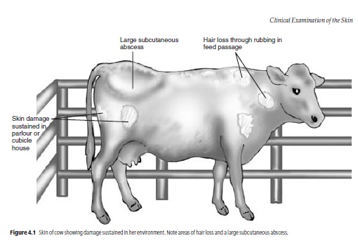
The environment of modern cattle, especially the
dairy cow, contains many features that may damage
the skin. The cubicles, the parlour and the floor may
have abrasive surfaces or sharp corners that can
cause injury to the skin, often repeatedly. Such problems
in the environment are especially likely to be
important if a number of cattle in the herd are seen
with identical superficial injuries. Overcrowding
and insufficient feeding facilities may also contribute
to poor coat condition including superficial skin
damage (Fig. 4.1).
Abnormalities such as a very poor coat, evidence
of excessive self-grooming or large areas of alopecia
may be seen from a distance, but the areas of the
skin must be closely examined too. Opportunities
to examine the skin arise as each part of the body is
examined, but in order to get a general impression
of the skin it can be assessed separately before the
more detailed examination of each area begins.
Visual appraisal of the skin
The whole body surface is methodically inspected
initially from a distance and then more closely, looking
for areas of abnormal skin or hair which will later
be subjected to closer scrutiny. Healthy animals have
lick marks on their skin, especially over the flank
and shoulders. Pruritus, for example that caused by
heavy louse infestation, may cause excessive grooming
and the presence of more lick marks than normal.
Repeated rubbing can lead to hair loss and thickening
of the skin. The presence of any obvious abnormalities,
including swellings or discharging abscesses,
should be noted for further investigation
later. Damp areas caused by sweating may be seen in
pyrexic animals and in warm weather. Skin loss
through injury may be seen. Gangrenous changes in
the skin and deeper tissue may have arisen through
loss of circulation and may be seen or noted during
manual appraisal of the skin.
Manual appraisal of the skin
This should involve as much of the body surface
as possible, using caution when touching sensitive
areas which might cause the animal to kick. Manual
appraisal will enable the clinician to detect lesions
which are not immediately visible, for example beneath
matted hair. Any abnormalities detected are
subjected to further scrutiny which may necessitate
removal of hair and examination of the skin in good
light with the aid of a hand lens. Enlargement of
lymph nodes may be detected at this stage (see
below). The thickness of the skin and the presence of
any subcutaneous oedema or infection should also
be noted. The average skin thickness in adult cattle is
6 mm, with decreasing thickness being evident from
the dorsal to the ventral body surfaces. The skin over
the brisket is quite thick and mobile. This area of skin
may have a spongy texture when compressed and
may give an impression of subcutaneous oedema although
it does not pit on pressure. Genuine oedema
which does pit on pressure may be seen in this area
and between the mandibles in cases of right sided
cardiac failure. The skin covering the lower limbs is
relatively immobile.
Manual examination of the skin will also allow assessment
of skin turgor – its resilience and flexibility.
Picking up a skin fold between finger and thumb and
releasing it provides a general assessment of the animal’s
state of hydration. In a well hydrated animal
the pinched skin falls immediately back into place; in
a dehydrated animal the return to normal is delayed.
The best site for this test is the skin of the upper
eyelid.
Pathological thickening of the skin occurs in a number
of skin conditions, including sarcoptic mange. Thickening
in the form of callus formation can occur in areas
of skin, including those covering joints, which are
repeatedly subjected to trauma. Examples include
the elbows and hocks in animals with poor bedding.
Distribution of skin lesions
This is of diagnostic importance. Lesions caused by
photosensitisation are commonly seen in lightly pigmented
areas on the dorsal parts of the body which
are exposed to sunlight. Such lesions are not normally
seen in pigmented areas. Ringworm lesions
in calves are particularly common on the head and
neck, but also occur elsewhere.
Description of the skin lesions
The clinician should try to determine exactly what
abnormalities are present in the skin, which tissues
are involved and how deeply the disease process
extends into and over the skin. The larger external
parasites such as lice may be seen at this stage. Skin
temperature, thickness, consistency and colour are
observed and compared with adjacent areas. The
presence of subcutaneous oedema or increased skin
turgor is noted: these abnormalities may be caused
by hypoproteinaemia or heart failure and dehydration,
respectively, but they can also be the result of
local pathology. When numbers of skin lesions are
found it is important to determine if they share the
same aetiology. They may represent different stages
of one disease process. More than one condition can
be present at the same time.
There may be abnormalities in the sebaceous and
sweat glands or gross proliferation of the superficial
layers. Self-inflicted trauma can greatly modify and
mask the clinical picture. Skin abnormalities may
involve some or all of the component structures of
the skin: the hair, follicles, epidermal, dermal and
subcutaneous tissues.

Clinical Examination of the Lymphatic System


 Lymphatic system
The lymphatic system consists of the carcase lymph
nodes, the network of lymph vessels which connect
them and the lymphatic parts of the spleen. Many of
the nodes are readily palpable in the healthy animal.
Others can be palpated only when enlarged. Details
of the location of the individual nodes and their ease
of palpation are given below. The lymph vessels are
normally palpable only if they are enlarged. Some
vessels may be seen and palpated as they run subcutaneously
towards the regional lymph nodes.
Clinical examination of the
lymphatic system
Grossly enlarged lymph nodes may have been seen
during observation of the patient before it is handled.
Further observation and palpation is possible when
the animal is restrained. The lymph nodes can be examined
as a separate system or checked during the
examination of the skin when the clinician’s hands
run over the whole body surface. Each paired node
should be compared for size and consistency with
the contralateral node.
Lymph node enlargement
This may occur for two main reasons.
(1) Enlargement of one or more lymph nodes may occur
in cases of infection of the lymphatic system. This
can occur in a number of diseases including
bovine tuberculosis, actinobacillosis and a number
of other bacterial infections. It can also occur
in both forms of bovine leucosis – enzootic bovine
leucosis (EBL) and sporadic bovine leucosis. EBL is
an uncommon but notifiable disease in the UK.
Infection is widespread in some other countries.
Any animal over 2 years of age with enlargement
of the carcase lymph nodes and in which bovine
leucosis is suspected is blood tested for serological
evidence of EBL. Positive cases of EBL are
slaughtered.
Cases of sporadic bovine leucosis may be examined
further to determine which carcase and palpable
visceral lymph nodes are involved. Gross
lymph node enlargement may be seen, for example,
in the prescapular lymph nodes. In most
cases some enlargement is present in other
lymph nodes. Ulceration of affected lymph
nodes may occur. Areas of tumour tissue may be
seen in the skin and in the thymus. Internal lymphoid
tumours may be found in many locations
including the heart base, the mediastinum and
wherever lymph nodes are present. Affected
lymph nodes are usually non-painful to the
touch but may interfere with many body functions.
Heart base and thymic tumours may
obstruct venous return. Mediastinal tumours
may compress the oesophagus causing bloat or
dysphagia.
(2) Lymph node enlargement in response to local infection
or inflammation in the region of the body drained
by the lymph node involved. In these circumstances
the lymph node is acting as a sentinel of
local disease. The enlarged node may be warm
and inflamed, and sensitive to the touch. On finding
an enlarged lymph node the clinician should
examine the area draining into the affected node
for evidence of any pathological problem. As
with tumour infiltration, the enlarged lymph
nodes may affect the function of adjacent organs.
Location of the carcase lymph nodes
Many of the nodes are paired and should be compared
for size and consistency. Lymph nodes are
normally firmer than adjacent muscle and other soft
tissues (Fig. 3.1).
Submandibular lymph nodes
These are situated and are palpable on the medial
aspect of the ‘angle of the jaw’ where the horizontal
and vertical rami of the mandible meet. Normal size
is 1.5 to 2cm at maximum diameter.
Parotid lymph nodes
Often these are not palpable unless they are enlarged
through local infection or tumour formation. These
small nodes lie subcutaneously just below the temperomandibular
joint. Normal size is 0.5cm.
Retropharyngeal lymph nodes
These nodes lie in the midline dorsal to the pharynx.
If enlarged they can be palpated by placing two
fingers of one hand on either side of the larynx. The
fingers of the two hands are advanced towards each
other just dorsal to the larynx. In normal animals the
retropharyngeal nodes are rarely palpable and it is
possible to advance the fingers (as described above)
towards each other until they are separated only
by the compressed pharynx. Dysphagia and dyspnoea
with stertorous breathing may be seen in
animals in which the retropharyngeal nodes are enlarged.
The nodes may be up to 4 cm in diameter
when enlarged.
Prescapular lymph nodes
These nodes lie subcutaneously and underneath the
cutaneous muscle just anterior to the shoulder joint.
It is often possible to palpate them directly in front
of the shoulder. They may also be reliably located by
extending the fingers and pressing them forward
from the shoulder joint onto the neck. The fingers
push against the prescapular node even if it is small,
thus identifying its position. Further advance causes
the fingers to rise over the node and down onto the
neck in front of it, thus obtaining an estimate of
the size of the node. The prescapular nodes vary in
size and may be small and round or elongated in
a dorsoventral direction. Normal size in adult is
1cm¥ 3.5cm.

vendredi 10 juin 2011

LES DEUX MODES DE REGULATION DE LA CONSOMMATION VOLONTAIRE


La consommation volontaire de la vache laitière subit une double régulation, d'abord volumétrique puis biochimique permettant un bon ajustement aux besoins énergétiques comme chez tous les animaux.

• La régulation volumétrique met en cause l'encombrement des préestomacs qui est directement lié à la faible densité énergétique des aliments fibreux (UFL par kg MS ou par 1) et à leur lente dissolution ruminale, et qui est quelque peu en relation inverse avec leur digestibilité.

Elle restreint le niveau d'ingestion des rations à base de fourrages, d'autant plus que ceux-ci sont plus médiocres.

Réciproquement, l'amélioration de la qualité des fourrages et au besoin l'association d'un minimum de concentrés équilibrés pour augmenter la concentration nutritive a le triple mérite d'accroître conjointement l'ingestibilité, la digestibilité et le rendement métabolique.

A ce titre, un optimum de concentration nutritive, autorisant une ingestion maximale, est atteint quand se trouvent couverts les besoins nutritifs. Il se situe vers 67 à 73 p. 100 de digestibilité, 15 à 17 p. 100 de cellulose brute (19 à 21 p. 100 d'ADF, 25 à 28 p. 100 de NDF) et 60 à 70 p. 100 de concentrés dans la ration totale.

Évidemment, il tend à s'élever avec le niveau de production,

• Au-delà de ce seuil, correspondant au point d'inversion du mode de régulation de l'ingestion volontaire, l'encombrement de la ration ayant cessé d'être limitant, c'est la régulation biochimique qui intervient pour égaliser la prise énergétique aux besoins.

Cet ajustement de la consommation volontaire en fonction des besoins énergétiques est commandée par le niveau de résorption des acides gras volatils.

Il peut être entravé par l'excès d'acide lactique (acidose) ou de corps cétoniques (cétose).

Il dépend de la demande énergétique de l'organisme, notamment de sa productivité, elle-même tributaire de l'efficacité des apports protéiques (voir figure 40).

Si la concentration énergétique (UFL/kgMS) augmente plus que nécessaire.

l'animal réduit son niveau d'ingestion (en kgMS) pour un même apport d'UFL : il se manifeste donc une totale substitution des concentrés à la place des fourrages sur une base énergétique. Il en résulte une aggravation du prix de revient de la ration (dans la mesure où l'UFL de concentrés coûte plus cher que l'UFL de fourrage).

Il en dérive aussi une certaine déviation du partage de l'énergie nette en faveur de l'adipogenèse car l'accroissement de la proportion alimentaire de concentré élève la proportion propionate/acétate dans le mélange des acides gras issus des fermentations ruminales.

L'exagération de l'apport de concentré expose ensuite à une acidose digestive dont une des premières conséquences est

mercredi 8 juin 2011

ACCUMULATION EXCESSIVE D'AMMONIAC DANS LE RUMEN


Une dangereuse accumulation rumi-nale d'ammoniac résulte d'une dysharmonie entre une ammoniogenèse à tendance explosive et une protéo-synthèse microbienne toujours relativement lente et limitée.

• L'ammoniogenèse trop rapideest encore renforcée par des apports trop abondants, trop brutaux ou trop peu fractionnés d'azote trop rapide-ment dégradable ;

• La protéosynthèse microbienne trop faible est davantage ralenlie par une inadaptation de la microflore ou un approvisionnement de glucides fermentcscibles insuffisant, désynchronisé ou discontinu par rapport à la libération en ammoniac (voir figure 36).

Les manifestations suraiguës en sont bien connues en raison de leur caractère spectaculaire : tétanie de nutritionet tétanie d'herbage (compliquée d'hypomagnésémie), ainsi que enté-rotoxémies vraies (par prolifération ruminale de Clostridiales).

Les formes cbroniques, beaucoup plus insidieuses et polymorphes sont largement sous-estimées alors qu'elles sont en fait beaucoup plus graves économiquement :

- "Fièvres de lait" classiques ou atypiques (syndrome de la vache rampante ou couchée) ;

- Gélose, infertilité. mortalité em-bryonnaire et avortement (surtout chez les génisses, vraisemblablement par le fait qu'à la différence des vaches en lactation, elles ne peuvent profiter de l'émonctoire mammaire) ;

- Altération de la qualité de la corne podale, fourbures ;


- Inflammation mammaire et baisse générale de l'immunité 
favorisant le déclenchement de mammites (voir figure 37).


La digestion azotée dans le rumen passe par le stade critique de l'ammoniac dont l'accumulation expose à de multiples conséquences pathologiques : l'alcalose chronique ou même une intoxication aiguë telle que la tétanie de nutrition ou d'herbage. La prévention consiste d'abord à atténuer l'ammoniogenèse par des apports azotés modérés, à dégradation progressive et en distribution fractionnée. Elle impose aussi à convertir cet ammoniac en protéines microbiennes par la fourniture suffisante, synchrone et bien répartie de glucides fermentescibles, avec le double bénéfice d'éviter la toxicité ammoniacale et d'améliorer le rationnement azoté.

mercredi 4 mai 2011

INTERET DE L'APPORT DE PROTÉINES INDÉGRADABLES


Les PDIM ne pouvant couvrir à elles seules, en plus de l'entretien, qu'une production maximale de 12 à 15 kg de lait par vache et par jour, les VLHP doivent recevoir en complément des sources de PDIA.
Ces protéines indégradables (dans le rumen), sont nécessairement bien digestibles (clans l'intestin grêle) et fort bien pourvues en acides aminés indispensables.
En effet, elle doivent constituer un ensemble équilibré avec les PDIM, en vue de soutenir la protéo-synthèse mammaire, de manière d'autant plus obligatoire et rigoureuse que celle-ci est plus intense. Les protéines microbiennes sont relativement déficientes en méthionine. 
De ce fait, les sources de protéines indégradables riches en méthionine sont intéressantes, telles que les drêches de brasserie ou le gluten d'amidonnerie.
En outre, celui-ci contient en abondance de la proline qui permettrait une épargne métabolique d'arginine à laquelle on attribue un effet favorable sur la synthèse de somatotropine et donc sur la production laitière.
De façon générale, chez les VLHP, on a intérêt à utiliser des protéines indégradables de qualité, telles que le tourteau de soja (qui gagne à être amélioré pour des ruminants grâce à un léger surtraitement thermique à 130° C), foin de luzerne ou farine de luzerne déshydratée (dont les protéines ont été insolubilisées au cours du séchage).
En raison des risques potentiels de transmission de l'Encé-phalopathie Spongiforme Bovine (BSE) ou "maladie de la vache folle",
l'emploi de farines animales est à présent interdit chez les ruminants.
Une augmentation mesurée de l'indé- gradabilité protéique peut être obtenue par des traitements spécifiques ménagés, soit par des acides, soit surtout par le formol (solution de for-maldéhyde, notamment selon le procédé de l'IXRA), soit encore par les tanins (par exemple en utilisant les "eaux blanches" dérivées de la technologie du bois de châtaignier aggloméré, (voir figure 35).
Pour parfaire la supplémentation azotée chez les VLHP en début de lactation (DL), il est aussi possible de recourir aux acides aminés de synthèse, sous forme protégée vis-à-vis de la dégradation microbienne rumi-nale par polymérisation ou enrobage ; en milieu acide, dans la caillette, ils sont prévus pour redevenir disponibles. Comme les déficits résiduels sont habituellement multiples, il faut alors obligatoirement associer au moins la lysine (de l'ordre de 25 à 30 g/VL/j) et la méthionine (environ 15 g/VL/j), sinon la phénylalanine, la thréonine, la valine... Ainsi, l'alimentation azotée des VLHP-DL retrouve les mêmes règles de l'équilibre des acides aminés indispensables que chez les monogastriques.